Carotid Plaque Imaging Enables Improved Carotid Artery Management
Carotid plaque imaging is changing carotid artery management because it lets clinicians study the plaque itself, not just
 Written and medically reviewed byDr. Abu BakarContributing writer · PharmD, PhD (Pharmacology)
June 14, 2026 · 8 min read
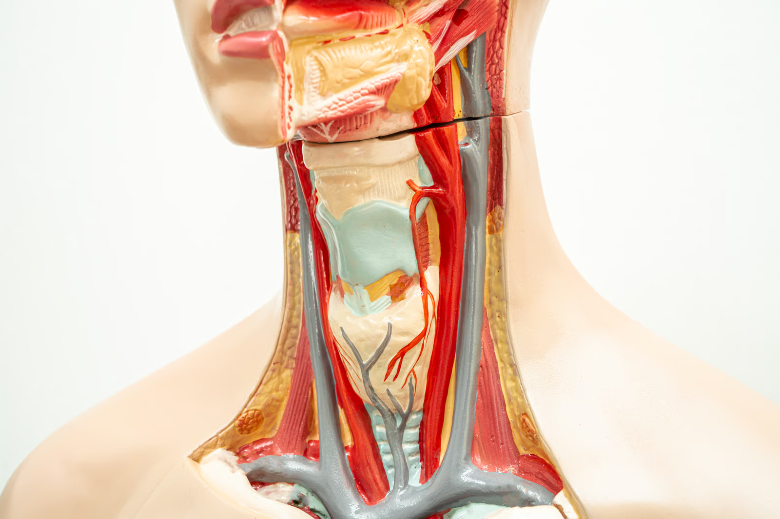
Carotid plaque imaging is changing carotid artery management because it lets clinicians study the plaque itself, not just the size of the blockage. This is a critical step towards stroke prevention. Traditionally, clinical decisions regarding carotid arteries have been based primarily upon the presence of luminal stenosis; however, it is now evident that intraplaque hemorrhage, lipid core, thin/ruptured fibrous cap, ulceration, and inflammation are better predictors of plaque instability and subsequent embolic complications. Using duplex ultrasound, CT angiography, magnetic resonance imaging, and even molecular imaging, clinicians can create an informed profile and base their decisions on whether to use medical treatment, close monitoring, surgical intervention, or stenting of the carotid arteries.
Why It Matters
Stroke prevention depends on finding the dangerous lesion before it causes harm. Stroke is one of the foremost causes of death and disability worldwide; thus, it should be no wonder that treatment options for carotid diseases have traditionally hinged on clinical queries with immediate answers: Is the patient symptomatic, and what is the stenotic burden? The current top international guidelines continue to formulate their revascularization decisions along these lines, which are based on the foundation of evidence that underpinned the evolution of contemporary carotid care. However, this approach only addresses part of the biological story.
The problem is that two plaques with the same degree of stenosis may not carry the same stroke risk. A plaque that contains intraplaque hemorrhage, ulceration, or a thin/ruptured cap can behave very differently from a more fibrotic or heavily calcified lesion. In a patient level meta analysis, MRI detected intraplaque hemorrhage independently increased ipsilateral stroke risk in both symptomatic and asymptomatic carotid stenosis and outperformed many traditional clinical risk markers. Separate studies and meta analyses have also linked ulceration and MRI defined vulnerable plaque features with symptomatic presentation and future cerebrovascular events, including in lesions that are not severely stenotic.
This is why plaque imaging is important: it helps place biological information on a clinical anatomic finding. While duplex ultrasound will always be the preferred choice for initial evaluation due to its availability and familiarity in the assessment of both plaque presence and stenosis, CTA is promising thanks to its speed, availability, lack of operator dependence, and ability to delineate lumen, calcification, and many surface characteristics. MRI becomes particularly useful for the characterization of soft tissue components in plaque, providing excellent performance in areas like intraplaque hemorrhage and other markers of instability. There is also potential for molecular imaging technologies like PET in the detection of inflammation.
Who It Affects
Patients with recent neurologic symptoms are the first group likely to benefit from this shift. People who present with a transient ischemic attack, minor ischemic stroke, amaurosis fugax, or other focal ischemic symptoms often move quickly into a carotid workup. In these patients, plaque imaging can help explain why a modest or moderate stenosis still behaved like a dangerous lesion. Studies in symptomatic carotid disease have shown that vulnerable plaque features, including intraplaque hemorrhage, may remain clinically important even when the degree of stenosis is below the classic high-grade thresholds.
Asymptomatic patients may also be managed differently as plaque imaging becomes more common. Not every incidental carotid plaque needs surgery, and modern carotid care is increasingly careful about avoiding procedures that offer little net benefit. But this is exactly where better risk stratification matters. A major meta-analysis in asymptomatic carotid stenosis found that high-risk plaques were common and associated with a higher annual rate of ipsilateral ischemic events, which supports moving beyond stenosis percentage alone when deciding who needs closer surveillance and who should at least have a specialist discussion.
Clinicians across several specialties will need to speak the same imaging language. Neurologists, stroke physicians, vascular surgeons, interventionalists, radiologists, and primary care clinicians will all see the downstream effects of this change. The more imaging moves beyond “50%,” “70%,” or “80%” stenosis and into plaque composition, the more important standardized reporting becomes. Consensus work in the field has emphasized the limits of lumen-only imaging, and newer structured systems such as Plaque-RADS are designed to make reports more consistent and more clinically useful across modalities and centers.
Health systems will feel the pressure in workflow, training, and access. High-resolution vessel wall MRI is not yet plug-and-play everywhere, and some plaque MRI approaches have not been fully integrated into standard practice because dedicated equipment and expertise are not universally available. CTA is easier to deploy in many hospitals, but even then, advanced plaque assessment still depends on protocol quality and reader confidence. In that setting, unequal access becomes a practical concern, especially because stroke already carries a large burden in health systems worldwide.
Patients themselves will also need better explanations, not just better scans. A more detailed imaging result can improve care, but it can also create confusion if the report sounds alarming without clear context. Patients have to understand that a “high-risk” plaque feature does not automatically mean surgery, and a “stable” plaque does not mean the artery can be ignored. The value of plaque imaging is greatest when it supports shared decision-making rather than replacing it.
What Changes
The diagnostic pathway will become more layered and more selective. In many centers, duplex ultrasound will remain the starting point because it is still the most practical first-line test for carotid assessment. The next step, however, is increasingly tailored to the clinical question. CTA is useful when clinicians need a fast, widely available view of stenosis, calcification, and plaque surface detail. MRI becomes especially helpful when the question is whether the plaque contains hemorrhage, lipid-rich material, or cap disruption. In optimized protocols, vessel wall MRI can deliver this richer tissue detail, although it requires time, protocol control, and experienced interpretation.
Patient selection for surgery or stenting will likely become more nuanced, especially in gray-zone cases. Established guideline-based care is not disappearing: symptomatic 50% to 99% stenosis and selected severe asymptomatic stenosis still anchor current revascularization decisions. What plaque imaging adds is a sharper lens for the uncertain cases. A patient with moderate stenosis and convincing plaque vulnerability may deserve earlier specialist review, while a patient with asymptomatic disease and no high-risk plaque features may be a stronger candidate for careful medical management and surveillance. In other words, plaque imaging is most useful when the decision is not already obvious.
The best medicine remains key to any treatment plan, and imaging could possibly improve the targeting of such medication. While the latest advancements in the treatment of carotid artery disease have been centered largely around the importance of lipids reduction, antiplatelet medications, where appropriate, blood pressure control, diabetes control, and smoking cessation, the newer carotid guidelines outline specifically how to prescribe antiplatelets and lipids for symptomatic and asymptomatic disease, indicating that imaging does not replace preventive measures. Instead, what imaging offers is an insight into identifying those patients that would require closer follow up and revascularization considerations.
The process of imaging should be made systematic and the use of multidisciplinary review should become routine. One of the main reasons why plaque imaging has not been easy to implement is due to the different protocols and methods that have been applied by various centers. The use of a systematic framework ensures that a clinically useful image is produced from the technical image. The use of Plaque RADS becomes critical in this case since it is created specifically as a multimodality language and not just for one modality.
But there are true limitations, and those should be stated explicitly. Improved imaging sensitivity will produce more abnormal findings, yet not every abnormal finding necessarily warrants aggressive therapy. There is not even universal agreement on appropriate management levels of certain plaque characteristics, and the consensus statements of experts keep emphasizing the necessity of randomized clinical trials establishing the value of plaque imaging before its impact on standard care paths becomes decisive. It is essential not to get carried away with what is seen on the screen, despite its importance.
Future tools may make plaque imaging more consistent, but they will need careful validation. Systematic reviews suggest that AI and machine learning models show promising diagnostic performance for classifying carotid plaque vulnerability, and current research is also pushing molecular imaging toward better visualization of plaque inflammation and other biologic activity. Those developments are encouraging because they could reduce reader variability and improve risk prediction. Still, the evidence base remains heterogeneous, and newer tools should be seen as complements to expert clinical judgment, not replacements for it.
Taken together, carotid plaque imaging offers a more precise way to think about stroke prevention. It helps clinicians move beyond a narrow, percentage-based view of disease and toward a model that reflects plaque biology, patient context, and evolving evidence. That does not mean every center must suddenly abandon lumen-based practice. It means carotid care is becoming more individualized, and the most useful question is no longer only “How tight is the stenosis?” but also “How dangerous is this plaque, for this patient, right now?”
References
- Dakis K, Nana P, Chaidoulis A, Spanos K, Batzalexis K, Giannoukas A, et al. Carotid Plaque Vulnerability Diagnosis by CTA versus MRA: A Systematic Review. Diagnostics (Basel). 2023;13(4):646. Direct URL: https://pmc.ncbi.nlm.nih.gov/articles/PMC9955971/ [\[pmc.ncbi.nlm.nih.gov\]](https://pmc.ncbi.nlm.nih.gov/articles/PMC9955971/)
- Saba L, Cau R, Murgia A, Nicolaides AN, Wintermark M, Castillo M, et al. Carotid Plaque-RADS: A Novel Stroke Risk Classification System. JACC: Cardiovascular Imaging. 2024;17(1):62-75. Direct URL: https://doi.org/10.1016/j.jcmg.2023.09.005 [\[radiology….ornell.edu\]](https://radiology.weill.cornell.edu/research/publications/carotid-plaque-rads-novel-stroke-risk-classification-system)
- Naylor AR, Rantner B, Ancetti S, de Borst GJ, De Carlo M, Halliday A, et al. European Society for Vascular Surgery (ESVS) 2023 Clinical Practice Guidelines on the Management of Atherosclerotic Carotid and Vertebral Artery Disease. European Journal of Vascular and Endovascular Surgery. 2022. Direct URL: https://doi.org/10.1016/j.ejvs.2022.04.011 [\[forschung.h-och.ch\]](https://forschung.h-och.ch/en/publikationen/11763-european-society-for-vascular-surgery-esvs-2023-clinical-practice-guidelines-on-the-management-of-atherosclerotic-carotid-and-vertebral-artery-disease)
- Jie P, Fan M, Zhang H, Wang O, Lv J, Liu Y, et al. Diagnostic value of artificial intelligence-assisted CTA for the assessment of atherosclerosis plaque: a systematic review and meta-analysis. Frontiers in Cardiovascular Medicine. 2024;11. Direct URL: https://www.frontiersin.org/journals/cardiovascular-medicine/articles/10.3389/fcvm.2024.1398963/full [\[frontiersin.org\]](https://www.frontiersin.org/journals/cardiovascular-medicine/articles/10.3389/fcvm.2024.1398963/full)
One story a day
The story of the day, in your inbox
One health journey each morning — no advice, no alarm, just company for the road.