Clinician Adoption of AI to Detect LV Systolic Dysfunction in Everyday Practice
Left ventricular systolic dysfunction is the heart problem behind many cases of heart failure, and it often develops
 Written and medically reviewed byDr. Abu BakarContributing writer · PharmD, PhD (Pharmacology)
June 19, 2026 · 7 min read
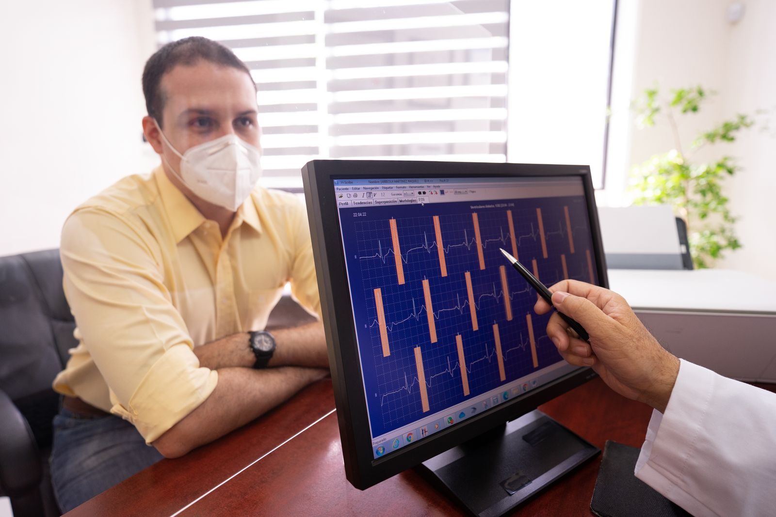
Left ventricular systolic dysfunction is the heart problem behind many cases of heart failure, and it often develops quietly before a patient ever feels shortness of breath, swollen or unusually tired. That is why clinicians are watching new artificial intelligence tools so closely. These systems can analyze tests that are already part of routine care, especially the 12-lead ECG and low-cost bedside ultrasound and raise an early flag when the heart’s pumping function may be reduced. The promise is practical rather than futuristic because it allows clinicians to catch risk earlier, confirm it with the appropriate follow-up test and begin treatment before the patient reaches a crisis point.
Why It Matters
Left ventricular systolic dysfunction means the heart’s main pumping chamber is not squeezing strongly enough to move blood forward efficiently. This condition can occur well ahead of any manifestation of heart failure without drawing attention, but once the symptoms appear, the outcomes will certainly be serious for both the patient and the healthcare system. Millions of adults experience heart failure annually in the US alone, with many dying from this condition every year. The treatment of such patients imposes a huge financial burden in terms of hospitalization, medication and ongoing follow up visits. In a broader perspective, heart disease is currently the leading cause of mortality globally. The earlier window becomes significant because it is not merely a diagnosis that appears in an echo cardiogram report. It is a phase during the course of a disease process wherein medical professionals can intervene and prevent recurrent decompensation, fluid overload, and potentially avoid hospitalization altogether. Asymptomatic left ventricular systolic dysfunction has been described in medical literature as pre heart failure and heart failure guidelines today place great importance on preventing and early detection of heart failure. At the same time, routine echocardiography for entire populations has not been recommended as a practical screening strategy, which leaves clinicians needing a lower cost, more scalable way to decide who really needs formal imaging. Artificial intelligence offers that scalable option because it can pull more meaning from data clinicians already collect every day. In a large 2019 study, an AI-enabled ECG identified low ejection fraction with an area under the curve of 0.93, sensitivity of 86.3%, and specificity of 85.7% in an independent validation set of 52,870 patients. The same study also found that patients without documented ventricular dysfunction but with a positive AI screen had a much higher future risk of developing dysfunction. For clinicians, that changes the ECG from a test used mainly to assess rhythm and conduction into a possible early screening signal for structural heart disease The real world application increases the applicability of the results to clinical practice. As seen in the EAGLE pragmatic trial, where more than 22,000 adults with no previous heart failure were screened for low ejection fraction by means of routine care ECG and the clinicians who had access to AI diagnoses identified more patients with low ejection fraction within 90 days compared to those practicing under routine care, the overall use of echocardiogram across all participants remained relatively unchanged, while follow-up imaging significantly increased among the positive AI group. This is precisely the desired effect that is typically expected from a good screening procedure. Trust and fairness sit at the center of adoption. WHO guidance on AI for health emphasizes transparency, accountability, inclusiveness, and human oversight. That matters because clinicians are right to ask how a model performs in their own patients, not just in a research dataset. Recent work on ECG deep-learning models for heart failure prediction has shown that subgroup performance can differ, with weaker performance reported in some younger Black patient groups, especially young Black women. That does not erase the promise of AI screening, but it does mean hospitals need local validation, subgroup auditing, and ongoing monitoring if they want clinicians to rely on these tools in good faith
Who It Affects
Patients at risk of heart failure are the clearest group affected. In practice, that includes many older adults and people living with high blood pressure, diabetes, coronary artery disease, or a past heart attack. Some also carry a family history of cardiomyopathy or other heart disease. For these patients, an AI flag is not a diagnosis, but it can shorten the path from vague risk to the right confirmatory test. The practical value is simple: earlier recognition can open the door to earlier treatment decisions before the first major deterioration forces the issue.
Primary care clinicians will likely feel the biggest day-to-day effect. They manage risk factors over years, review routine ECGs, and often decide whether a subtle concern deserves a full cardiac workup. An AI prompt could help them notice a gradually developing problem that might otherwise be easy to miss in a busy clinic. But primary care is also where alert fatigue is most real. If the signal is not specific, interpretable, and tied to a clear next step, clinicians will ignore it. The EAGLE trial mattered partly because it tested AI-ECG in routine primary care rather than in an idealized research environment.
Cardiologists and diagnostic services will feel the downstream pressure. Better case-finding means more confirmatory echocardiograms, more referral questions, and more need to sort urgent follow-up from routine review. That can improve care if systems are ready. It can also clog imaging access if programs identify patients faster than services can confirm disease. This is where AI-guided point-of-care ultrasound may help by moving some initial assessment closer to the bedside, especially when nonexpert users can still achieve reasonable accuracy with AI assistance.
Payors, administrative, policy makers and patient advocates are included as well. Payors, administrative, governance, procurement and monitoring criteria all play an important role in reimbursement, staffing, economic modeling, and the actual process by which post-deployment performance is measured. Economic modeling of AI-ECG screening suggests that AI-ECG screening can be effective from a cost perspective where focused screening is concerned, although more general adoption depends on how many follow-up tests and visits result from the program. On the other hand, patient advocates will remain vocal regarding transparency, fairness, and communication of what the AI warning means.
What Changes
Earlier identification becomes more realistic, but only when the alert leads to action. Artificial Intelligence could detect potential signs of left ventricular systolic dysfunction through a routine ECG or ultrasound before there are any apparent symptoms, but the true advantage is seen only if that alert prompts further investigation through imaging, evaluation by the physician, and subsequent treatment in case of presence of the disease. In reality, the ideal incarnation of such technology will not be the most assertive one. It will be the silent one.
Workflow will be shifted towards triage and follow-up. The clinics adopting such technologies will require a straightforward process of addressing basic concerns like “who sees the alert?,” “who verifies the alert? who orders the echo? who tracks the task?, and who tells the patient?” This is exactly the reason why recent implementation studies focus on decision support embedded within EHRs. Clinician adoption tends to increase when the technology streamlines processes and offers a concrete plan of action; and it often fails when it introduces uncertainty or extra steps without an owner.
Resource pressure will also increase unless programs are designed carefully. Even a modest rise in true-positive detection can strain echo labs and cardiology access if a health system is already running at capacity. That is one reason targeted screening is likely to be more workable than a broad untargeted rollout. The economic literature points in that same direction: AI-enabled screening looks more attractive when used in focused populations and when follow-up is purposeful rather than diffuse. For clinicians, that means adoption will feel safer when the program is paired with realistic capacity planning from the start.
Clinician judgment will remain central. Most doctors are unlikely to treat AI output as a final diagnosis, and that is probably the right approach. In everyday practice, these technologies are useful mainly as decision aids and second opinions rather than as substitutes for history, signs, previous imaging, other diseases, and patient choice. Such a restrained application will undoubtedly delay widespread implementation, but it also reduces the risk of unintended consequences and enables physicians to feel more at ease in terms of their professional obligations. Eventually, success will be determined less by hype and more by evidence of reliable results and accountability.
The long-term shift is not just technical. It is organizational. Health systems that want AI detection of left ventricular systolic dysfunction to work will need subgroup audits, privacy safeguards, local validation, and regular checks for model drift as populations, devices, and clinical practice change. Clinicians do not need a flashy dashboard. They need a governed tool that helps them catch weak heart pumping earlier without adding waste, delay, or inequity. That is the real adoption test. If the tool helps clinicians see more clearly and act more easily, it will spread. If it adds noise, it will not.
References
- Heidenreich PA, Bozkurt B, Aguilar D, Allen LA, Byun JJ, Colvin MM, et al. 2022 AHA/ACC/HFSA Guideline for the Management of Heart Failure: A Report of the American College of Cardiology/American Heart Association Joint Committee on Clinical Practice Guidelines. Circulation. 2022;145(18):e895-e1032. Direct link [\[pubmed.ncb…lm.nih.gov\]](https://pubmed.ncbi.nlm.nih.gov/35363499/)
- Attia ZI, Kapa S, Lopez-Jimenez F, McKie PM, Ladewig DJ, Satam G, et al. Screening for cardiac contractile dysfunction using an artificial intelligence-enabled electrocardiogram. Nature Medicine. 2019;25(1):70-74. Direct link [\[europepmc.org\]](https://europepmc.org/article/MED/30617318)
- Yao X, Rushlow DR, Inselman JW, McCoy RG, Thacher TD, Behnken EM, et al. Artificial intelligence-enabled electrocardiograms for identification of patients with low ejection fraction: a pragmatic, randomized clinical trial. Nature Medicine. 2021;27(5):815-819. Direct link [\[europepmc.org\]](https://europepmc.org/article/MED/33958795)
- World Health Organization. Ethics and governance of artificial intelligence for health: WHO guidance. Geneva: World Health Organization; 2021. Direct link [\[who.int\]](https://www.who.int/publications/i/item/9789240029200), [\[iris.who.int\]](https://iris.who.int/server/api/core/bitstreams/f780d926-4ae3-42ce-a6d6-e898a5562621/content)
One story a day
The story of the day, in your inbox
One health journey each morning — no advice, no alarm, just company for the road.