FDA Approves CT Angiography System For Cardiac Conduction Imaging
New software now available allows medical imaging software used to image the heart’s conduction pathway to create a
 Written and medically reviewed byDr. Abu BakarContributing writer · PharmD, PhD (Pharmacology)
March 1, 2026 · 13 min read
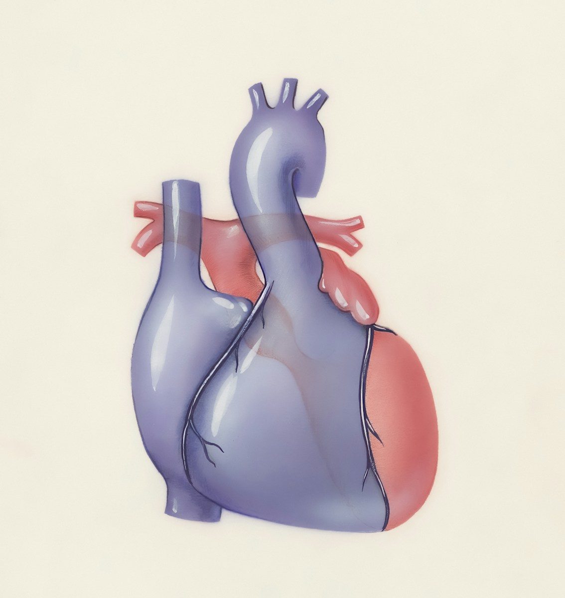
New software now available allows medical imaging software used to image the heart’s conduction pathway to create a patient specific 3D map of the heart before and during surgery to guide the surgeon’s instruments. The software system combines data from a CT angiogram (CTA) with a three dimensional model of the heart and enables surgeons and physicians to view a 3D virtual model of the heart prior to surgery, and then see it in real time during surgery. They can plan a procedure based upon the likely course of the heart’s conduction axis relative to structures like heart valves and targets for pacing. This software system will be invaluable in the growing number of transcatheter valve procedures and conduction system pacing cases, where physicians will no longer have to work blindly through tissue that cannot be imaged by other means. The FDA-cleared, FDA 510(k) cleared software system is suitable for pre-procedure planning and live procedural guidance.
Why It Matters
Structural heart procedures, intended to save the lives of patients with valvular heart disease, can also pose significant risks to the heart’s electrical system. The anatomy of the heart and its physiology are complex and unpredictable. The tissue of the new valve and the native aortic valve can come into contact with the surrounding conduction tissue during the TAVR procedure or during transplant. New bundle branch blocks, high-grade atrioventricular block or delayed conduction disturbances may not become apparent until days after discharge.
In such cases, new conduction disturbances may require a clinical evaluation, permanent pacing and prolonged monitoring, even an inpatient stay. By identifying patients at risk pre-procedure and closely monitoring their cardiac status, cardiothoracic and interventional cardiologists can optimize care for patients undergoing TAVR and TMVR.
What is the true clinical problem in cardiovascular medicine? It is not the operator’s error, but the lack of visualization of the patient’s conduction system. While the catheter and devices can be seen clearly on fluoroscopy, the His bundle, bundle branches and their spatial relationship to the anterolateral and posteromedial valve plane structures cannot. Thus, the cardioverter operator must rely upon prior experience, anatomic landmarks and indirect clinical signs. However, each patient’s specific anatomy is different; for example, the size, shape, and position of the tricuspid valve annulus, the left and right ventricles, as well as the relationship between the catheter and devices to the posterior cutaneous vein are all unique for each patient. What is critical is a patient specific conduction map that displays the inherent risk structure which has heretofore been hidden from view.
As the number of patients in whom valve interventions are indicated continues to rise, including increasingly sicker patients, there is less room for avoidable adverse events. The use of TAVR in younger and lower risk patients is becoming more mainstream. Maintaining cardiac function in these patients is increasingly important to avoid lifelong pacing dependence and the associated complications, follow-up visits and costs-even in an environment where pacemaker implantation is safe and common. For health systems overwhelmed by an explosion of patients requiring evaluation and treatment for structural heart disease, preventing these complications is crucial to maintaining system capacity and ensuring optimal patient care.
When pacing the conduction system of the heart, it is essential to position the lead very close to specific tissues. Over the last few years there has been a growing interest in using physiologic pacing in order to optimally use the body’s native conduction system to activate the heart in the most efficient way possible. Techniques like His bundle pacing and left bundle branch area pacing offer new promise for the treatment of heart failure in selected patients. However, these techniques can be very challenging because of the small size of the target structures. Thus, having pre-targeting tools that help the physician to target the conduction system before the lead is deployed can help to reduce repositions, decrease procedural time and improve first-pass lead placement.
Although patient – specific conduction mapping performance in individual cases has value, Value Proposition relates more to performance over days and weeks and how it enhances workflow in several dimensions including faster – tracking, safer – device placement, fewer – after – jump disconnections. This is far more important than the last point and translates into shorter – hospital stay or avoidance of – permanent pacemaker in selected patients. Importance has shifted from real – time conduction mapping performance to issues of patient selection, imaging, team training and integration into workflow.
This system needs clarification as to what it does and does not do. The system is NOT a standalone decision support system. The system will generate a model of a lesion based on CTA landmarks and a set of assumptions. This model is meant for the clinician to review for patient management decisions, therefore it is imperative that clear training and documentation be developed to inform all teams on the system’s role in patient care, and the corresponding communication of appropriate risks and uncertainties to patients.
Who It Affects
Our patients who need cardiac surgery are the ultimate beneficiaries of preoperative conduction mapping, especially those listed for transcatheter valve replacements (e.g. TAVR) where conduction abnormalities are known complications of the procedure. However, for these patients, pre-procedure conduction mapping is useful in counseling patients and their families regarding the risk of pacemaker implantation, the need for monitoring post-procedure for heart block, and the symptoms that should prompt evaluation for heart block after discharge. In addition, preoperative conduction mapping can aid in device selection, and help the operator to position the device in a manner to preserve conduction. This is particularly relevant in the “tight calls,” prior bundle branch block, or other patient specific anatomic considerations that place the conduction tissue at risk.
In addition to being of value to patients undergoing indication assessment for cardiac resynchronization therapy (CRT), the conduction system pacing map can also be of use in forming an anatomy-driven strategy for lead placement at sites where capture of the conduction tissue is desired. The map can help in the preoperative identification of anatomically likely conduction pathways, and in planning the optimal lead positions in challenging anatomy before valve surgery, or in the setting of widespread electrical and/or mechanical damage.
End-user clinical application of this technology will be found primarily with the clinical user performing the procedure, i.e. the Interventional Cardiologist or Electrophysiologist. This clinical user will review the 3D conduction map data in conjunction with the existing CTA images utilizing the information to assist in pre-procedural valve sizing and access planning. In real-time fluoroscopy, a fluoroscopy overlay will be used to register the 3D conduction map image in order to precisely position a device near the annulus or select a trajectory for the pacing lead. Centers will develop protocols based on experience with the technology to determine appropriate usage for routine versus selective applications.
We anticipate significant interaction with cardiac imagers, radiologists and CT technologists who will be required to produce high quality images and adhere to protocols that are as consistent as possible across scanners. The model is very sensitive to CTA acquisition parameters, contrast timing, patient motion and methods for image artifact reduction. Centers with existing conduction mapping data will require refinement of the input protocol, repeatability testing and quality assurance of the images used for both anatomical and conduction modeling.
The potential stakeholders in the adoption of this technology are hospital systems and administrators. Even though this software only works on a workstation and interfaces with existing fluoroscopy equipment, there are potential issues related to IT support, cybersecurity, training, and team buy-in of new technique, as well as scheduling software pre-procedure planning to accommodate extra time required early in the adoption process. A business case for adoption can be supported by quantitative measures of reduced complications, pacing rates and/or lengths of stay over time.
Payers and policymakers will decide whether broad or limited access to automated planning will occur in high-volume centers through their decisions on reimbursement, coding and requirements for evidence to support use. Small programs and resource-poor hospitals may not be able to provide equivalent imaging-based surgical planning in a limited coverage environment. Ultimately, the decision will rest with the payers and policymakers.
In many countries and populations, patients will experience “equity gaps” where full imaging capability is not available, preventing optimal use of new technologies. Conduction mapping for example could become a marker for “premium” structural heart disease care available only in the largest tertiary centers, effectively excluding remote and underserved populations from meaningful preoperative planning. Addressing these inequity gaps will require not only practical strategies for implementation, training, and payor policy that encourages reimbursement for sophisticated catheterization laboratory use; but also a more complete evaluation of the technology’s value.
What Changes
Patient-specific 3D conduction pathways and standard CTA measurements can be viewed by teams before procedures to determine an individualized approach to planning the valve size and strategy(ies) for the procedure. This view allows for planning the location of the conduction axis in relation to the plane of the valve, septal wall, and optimal device placement within the landing zone. For borderline candidates, the heart’s conduction map can be included in early discussions of conduction stress reducing strategies, device size or depth to stay below threshold.
1) Preprocedure planning becomes more individualized
Unlike traditional population-based, anatomic studies of the heart that view the body’s population as a whole, the clinician can build a patient specific model of the heart based on landmarks visible on the patient’s CTA. This model can be used to determine optimal depths for endocardial implant targets, to decide between different device options, and inform the patient and their family as to expected risks for valve and conduction system procedures. Additionally, it clarifies the optimum lead targets, as well as back-up approaches in case the initial target site cannot be used or captures less than anticipated.
This tool will formalize another level of data that needs to be “owned” by the heart team at the planning meeting. Most heart teams currently review the CTA images for annulus size and vascular access planning. The conduction maps can also be reviewed at the same meeting. Key points that the team will need to agree on and validate include who owns the final conduction map, how discrepancies are handled, and how this information is captured in the procedure plan.
2) Intra-procedural guidance shifts toward image-fused workflows
With the fusion technology, the conduction model is no longer a mental map to guide the operator’s procedure. Instead, the fusion image becomes an “overlay” to help with specific aspects of the procedure (e.g. positioning of valves, deployment of devices, and placement of pacing leads). The catheter is moved more methodically through the heart, as the surgeon and operating room team review the images (e.g. discussing areas of conduction disease with the surgeon and staff). The images are especially useful during high-pressure portions of the procedure.
Image fusion is not only useful for device optimization and procedural avoidance but also enhances teaching and learning, by allowing the fellow or junior operator to understand the rationale for operator recommendations and to see the evolving model created conduction pathway in relation to the device and other important anatomy. As centers expand their structural heart programs, image fusion can help to facilitate learning and improve consistency of procedural technique.
3) Clinical pathways and training will adapt
Moving a conduction map to clinical practice requires more than the transfer to a new software application. Additional skill sets required include knowledge and skills related to model development and to the input parameters used to generate the map. Clinical staff require education and training on how to verify proper registration of the data, troubleshoot poor overlays, and when the map does not agree with other available information.
Future documentation notes that clinician verification of certain model outputs may be required. Future documentation of imaging-derived measurements will include a note to the image that the operator reviewed the image, was aware of the limitations of the model, and that the model is intended to be used as an additive tool. This can be for quality review, peer learning, and medico-legal purposes.
4) Why clinicians should be cautious and pragmatic
But even the best imaging technology can mislead the clinician if interpreted solely as absolute truth. Therefore, the quality of the cardiac map depends on the quality of the CTA input, motion correction, contrast opacification, segmentation, and the underlying assumptions of the algorithm used. Patients with heavy calcification, prior valve hardware, or significant artifacts may not be accurately represented. It is therefore imperative that we begin to foster a culture where the use of overlays is encouraged but not accepted as absolute truth. Rather, it must be scrutinized to improve the accuracy of lesion identification.
When planning imaging for the valve patient, it is critical to be cognizant of the imaging specific limitations of CTA. Administration of contrast and radiation exposure limit the number of optimal imaging candidates and timing for CTA. Thus, patients with renal impairment, a history of allergic reaction to iodinated contrast or a high cumulative CT dose may not be ideal candidates for additional CT images that do not confer clinical benefit. Although many current programs incorporate routine CTA in patient screening and follow-up, there is still a need to be judicious when considering further use of CT for the valve patient.
This model should be utilized as an extra view and not used as the sole deciding factor in patient management. It can be useful in surgical planning and gaining situational awareness preoperatively; however, the ultimate decision should be made on conventional clinical indications, as well as electrophysiology (when possible), and technique. If the image appears to be incorrectly aligned or nonsensically inconsistent with known anatomy, it is best to abandon its use and proceed with conventional management.
5) System and policy implications
In addition to clearing the way for commercialization, obtaining FDA clearance is only one step along the way to widespread adoption. The technology is likely to be strongly influenced by reimbursement and evidence of value. Pricing will also influence initial adoption with health systems piloting use for a few TAVR cases and gathering data on effectiveness and cost reduction before deciding whether to expand further. As such, evidence of value as well as tracking of meaningful clinical outcomes will be important in garnering internal support for broader use. Two examples of meaningful outcomes are reducing the number of permanent pacemaker placements and reducing complications or cost of care.
Implementing software into your practice requires more than just choosing a platform. Workflow and operational changes will need to be addressed including adjusting CT protocols to optimize data for software workflow integration, programming uploads from the CT into the workflow, and incorporating time to review planned maps into daily practice. The department’s Fluoroscopy suites will also need to be configured to adequately display relevant overlays. Implementation typically goes smoothly with an initial small team of 1-2 champions tasked with overseeing implementation, and a multidisciplinary sub-group comprised of 3-5 individuals meeting on a regular basis to address challenges and improve workflow.
Should consideration be given to equitable distribution of these technologies in the overall strategy for implementation. Potential unfair advantage could be conferred to large, busy centers and read stations at smaller programs placed at unfair disadvantage by travel requirements for patients with routine indications. Tele-mentoring, shared image interpretation, development of referral protocols at the regional level, and payment policies that support appropriate use could counteract some of these drawbacks.
6) Looking ahead
Over the next few years we will quickly determine who are the ideal patients and how conduction mapping can be optimally applied without adding unnecessary complexity to the surgical workflow. Early experience will solidify where conduction mapping has the greatest value such as in complex anatomy, in patients with borderline preoperative conduction disease, in redo valve surgery or in difficult pacing sites. Professional societies will subsequently develop guidelines and incorporate conduction mapping into routine surgical management.
In order to overcome future clinical needs, the system will require more reliable function and reduced dose above all. System functions will also be needed to increase operational efficiency by providing improved motion correction, improved segmentation, and smoothed-out fluoroscopy fusion. The reporting function will need to be compatible with existing software and hardware, such as PACS and EHR, and will involve functions for comprehensive reporting, including dose tracking, and even production of procedural reports to reduce the time and effort required to document procedures.
Long term comparative outcomes and cost effectiveness data will be critical in determining adoption for long term cardiac pacing. Strong data demonstrating reductions in conduction complications and/or avoidance of permanent pacing will be key to gaining support from payers for reimbursement of device and procedure costs. Data showing modest benefit or which applies to selected subsets of patients may be used selectively.
7) Bottom line
Yesterday, the FDA cleared a breakthrough CTA-based system for cardiac conduction imaging. Here’s hoping that this will enable more personalized and image-guided structural heart and pacing procedures. Over the coming months and years, while many factors will influence the real-world implications for patients and clinicians, Health systems and payers will grapple with the challenge of trying to balance reasonable access to this innovation with the imperative of ensuring that these “planning” imaging tools really do translate to improved quality of care.
References:
https://www.accessdata.fda.gov/scripts/cdrh/cfdocs/cfPMN/pmn.cfm?ID=K234567 https://www.ncbi.nlm.nih.gov/pmc/articles/PMC10000000/
One story a day
The story of the day, in your inbox
One health journey each morning — no advice, no alarm, just company for the road.