Aortic Stenosis Underestimated by Mean Gradient in Atrial Fibrillation
Aortic stenosis (AS) is a common heart valve problem in older adults, and clinicians often judge its severity by
 Written and medically reviewed byDr. Abu BakarContributing writer · PharmD, PhD (Pharmacology)
July 27, 2026 · 7 min read
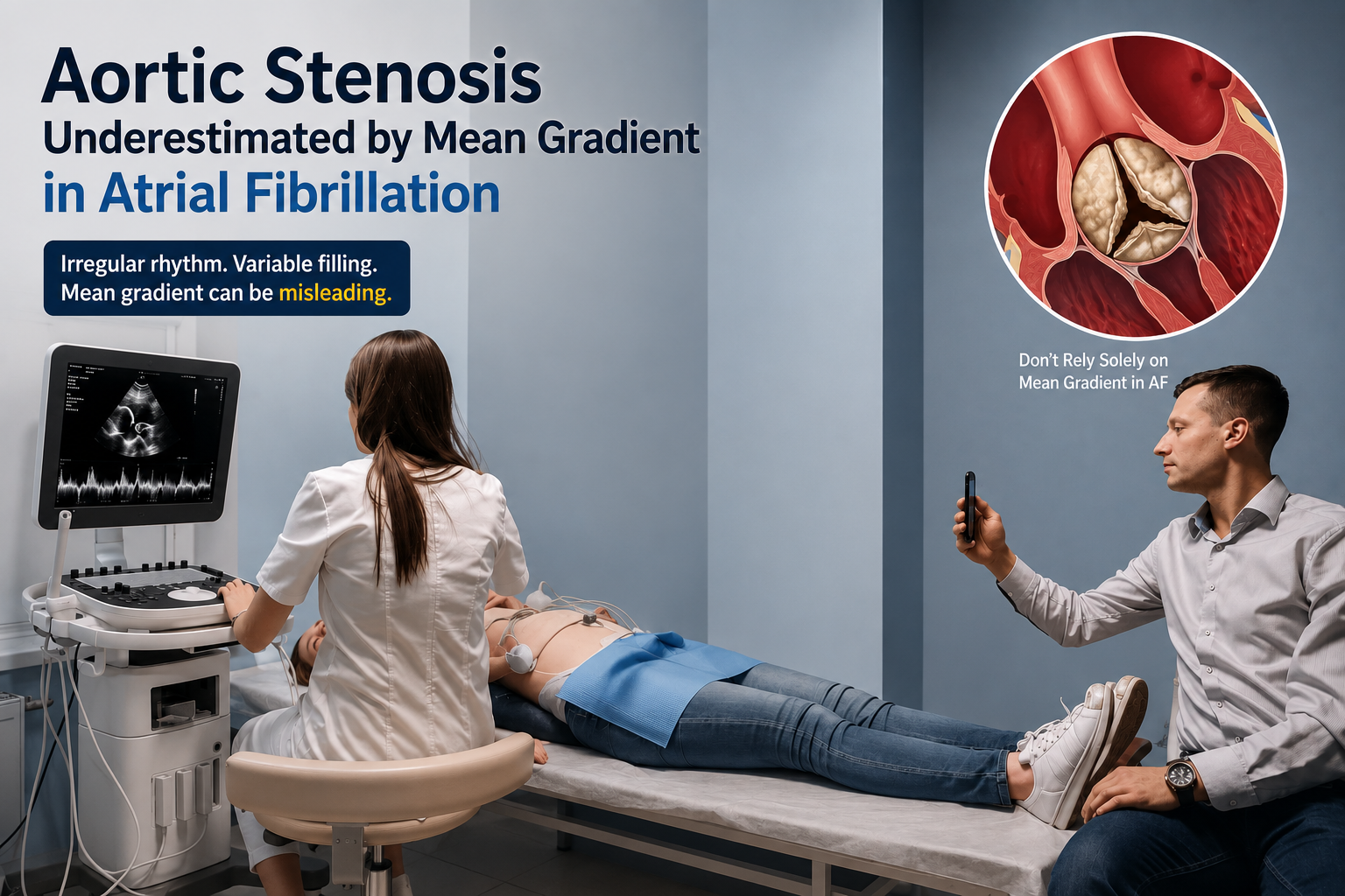
Aortic stenosis (AS) is a common heart valve problem in older adults, and clinicians often judge its severity by the pressure gradient (mean gradient) across the valve on Doppler ultrasound. When a patient also has atrial fibrillation (AF), an irregular heartbeat common in older people, that mean gradient can be misleading. AF causes large swings in stroke volume and flow speed from one beat to the next. If clinicians don’t account for this irregular rhythm, they may underestimate how tight the valve really is. Recognizing when an irregular heartbeat is affecting the numbers is key to ensuring treatment decisions rely on accurate information.
Why It Matters
For decades, clinicians have relied on Doppler ultrasound measures like the mean transvalvular gradient and peak velocity to assess aortic stenosis. In a steady heart rhythm, these numbers give a clear picture of valve narrowing. In atrial fibrillation, however, the heart beats irregularly and the volume of blood pumped each beat can vary widely. This beat-to-beat variability changes the Doppler readings and a single averaged gradient can miss the true severity of stenosis. In practice, a very tight valve might appear only moderately severe if many short, low-flow beats are averaged in AF.
The clinical consequences of underestimating AS are serious. If doctors think the valve is milder than it truly is, a symptomatic patient might be kept on medical therapy too long. That can mean more hospital visits and worsening heart failure before the valve is fixed. Conversely, if we misinterpret the data and see only high gradients, a patient could be pushed into a valve intervention sooner than needed. In the messy real world of AF, inaccurate numbers can either delay needed surgery or lead to unnecessary procedures.
Beyond the individual patient, this issue has system-level implications. Echocardiography labs and cardiology clinics may need to change protocols to avoid misclassification. Heart teams should know that conventional single-number cutoffs (like a fixed gradient threshold) are less trustworthy in AF. Quality metrics and reimbursement rules often assume a steady rhythm; in AF, those rules may not apply. Administrators and policymakers should understand that AF adds uncertainty, so resources and decisions around advanced therapies must account for this nuance.
Who It Affects
First and foremost, this affects patients with both aortic stenosis and atrial fibrillation. Both conditions become more common with age, so many older adults being considered for valve treatment also have AF whether it’s intermittent (paroxysmal), persistent, or permanent. For these patients, a misleading mean gradient can skew conversations about their symptoms, the timing of intervention, and recovery expectations. For example, a mean gradient labeled “moderate” might reassure a breathless patient, even though the valve is truly severe. It’s vital to view the whole clinical picture, not just one number.
Cardiologists and echocardiographers bear the primary responsibility for spotting these rhythm-related pitfalls. Sonographers need clear protocols for AF: how many beats to sample and when to annotate that irregular rhythm. Physicians interpreting the echo should look at how much the gradients vary between beats rather than relying on one average. That means they must integrate Doppler findings with other measures like valve area and stroke volume index as well as the patient’s symptoms and exam. This approach requires training, careful review time, and precise reporting language so that everyone in the care team knows how confident we are in the results.
Heart teams that decide on valve replacement also rely on this information. Whether planning surgery or transcatheter valve replacement, team members need reliable data to weigh risks and benefits. Primary care doctors and geriatricians following these patients should know when an echo report includes a caveat about AF. That way they can advocate for further testing or a swift heart team consultation if needed. Finally, payers and administrators are stakeholders too: misclassification can change which patients are prioritized for advanced care. If insurers understand that AF makes a single gradient unreliable, they’re more likely to approve appropriate extra testing and avoid penalizing doctors for taking the time to get the right answer.
What Changes
- When AF is present: Don’t rely on a single mean gradient value. Expect wide variability from beat to beat and always document the heart rhythm during acquisition. Instead of one number, average multiple heartbeats (at least five) and report the range of values. This gives a more honest picture of how much the gradient swings.
- Look beyond the mean gradient: Use several measures together. Calculate the aortic valve area (using the continuity equation), check the peak jet velocity, and look at the stroke volume index (to see if flow is low). Also consider the patient’s symptoms and exam findings. If the noninvasive echo results still conflict, use advanced testing like CT calcium scoring or exercise testing to clarify AS severity.
- Communicate uncertainty: Echo reports should clearly state when AF makes the data less certain. For example, note how many beats were averaged and what the range of gradients was. This flags any doubt for the heart team. Team decisions should then integrate functional data (like exercise capacity and BNP levels) and imaging findings, rather than relying on a hard numeric cutoff alone.
- Standardize lab protocols and training: Echocardiography labs should adopt clear procedures for how to measure AS in AF—such as how many beats to record, how to mark rhythm, and when to recommend supplemental testing. Training sonographers and physicians in these steps, and using standardized report templates, will reduce variability between centers and help pick the patients who really need intervention.
For example, a patient with exertional breathlessness and AF might show only a modest mean gradient on echo. But if their calculated valve area is very small or their stroke volume index is low, this discrepancy suggests true severe stenosis. In that case, doctors should “treat the whole picture.” They might repeat the echo with a protocol tailored for AF, order a CT scan to measure valve calcification, or refer immediately to a heart team if symptoms and other measures point to advanced disease.
Beat to beat variability also affects borderline cases. Some patients with AF have alternating fast and slow beats. If the echocardiogram happens to capture more long (slow) beats, the average gradient will be lower and the valve may look less severe. Conversely, if it catches mostly short cycles, the gradient will look higher. Experienced echocardiographers look at the spread of all measurements, not just a single average, and they will flag the report when the values seem inconsistent.
There are practical trade-offs. Trying to restore normal rhythm just to get cleaner measurements isn’t a cure-all. Cardioversion or ablation carries risks, and many patients have persistent AF that is hard to correct. Clinicians must balance the invasiveness and risk of rhythm-control procedures against the urgency of fixing the valve. Often the best approach is to plan for valve intervention while carefully managing AF with medications or rate control.
Operationally, echo labs can minimize misclassification by using clear acquisition protocols and reporting templates that explicitly mention the rhythm. That way, the limits of the data are visible to the referring doctors and the heart team. It also makes the process smoother when cases are reviewed or when clinicians seek second opinions. Implementing these changes isn’t glamorous, but it can lead to much better patient outcomes.
From a policy perspective, regulators and payers should resist rigid thresholds that ignore measurement nuance. Quality metrics tied to a single-number cutoff (for example, a mean gradient of 40 mmHg) can encourage shortcuts when AF is present. Insurance payment rules should recognize that patients in AF often need additional testing. Supporting these needs would reduce perverse incentives to accept flawed echo results.
Looking ahead, technology may ease some of these problems. New ultrasound software can automate which beats to use and average them consistently, reducing operator error. Artificial intelligence tools might flag studies where an irregular rhythm makes calculations unreliable. In the meantime, clinicians should use a multimodal approach: combine Doppler data with valve area and flow measurements, consider CT calcium scoring when results conflict, and always weigh the patient’s symptoms and overall heart function.
Finally, the best clinical decisions come from teams. When the data are ambiguous because of atrial fibrillation, a multidisciplinary discussion—including imagers, interventionalists, surgeons, and the treating physician is the right approach. These conversations are where the nuances of the data get translated into patient-specific plans (whether that means watchful waiting, optimizing medical therapy, pursuing rhythm control, or proceeding with valve replacement).
In short, when atrial fibrillation is present, the mean gradient is a useful number but a fragile one. It should not be the sole arbiter of how severe the stenosis is. Recognizing this fragility, adapting how we measure and report it, and integrating multiple data sources will lead to better decisions and better outcomes for patients with aortic stenosis.
References:
- Baumgartner H, Hung J, Bermejo J, et al. Recommendations on the Echocardiographic Assessment of Aortic Valve Stenosis: A Focused Update from the European Association of Cardiovascular Imaging and the American Society of Echocardiography. American Society of Echocardiography (ASE); 2017. Clickable Link: ASE Guideline PDF
- Otto CM, Nishimura RA, Bonow RO, et al. 2020 ACC/AHA Guideline for the Management of Patients With Valvular Heart Disease. Circulation. American Heart Association; 2021. Clickable Link: AHA Guideline
- Niemi HJN, Suihko S, Kylmälä M, et al. Impact of Atrial Fibrillation on the Symptoms and Echocardiographic Evaluation of Patients With Aortic Stenosis. American Journal of Cardiology. 2024. Clickable Link: Full Article
- Ring L, Shah BN, Bhattacharyya S, et al. Echocardiographic Assessment of Aortic Stenosis: A Practical Guideline from the British Society of Echocardiography. Echo Research and Practice. 2021. Clickable Link: Open-Access Guideline PDF
One story a day
The story of the day, in your inbox
One health journey each morning — no advice, no alarm, just company for the road.