FDA Clears Karl Storz Rubina Lens Exoscope for Open Surgery
The clearance of a camera based exoscope for open surgery adds a new tool to the surgical toolkit….
 Written and medically reviewed byDr. Abu BakarContributing writer · PharmD, PhD (Pharmacology)
July 3, 2026 · 8 min read
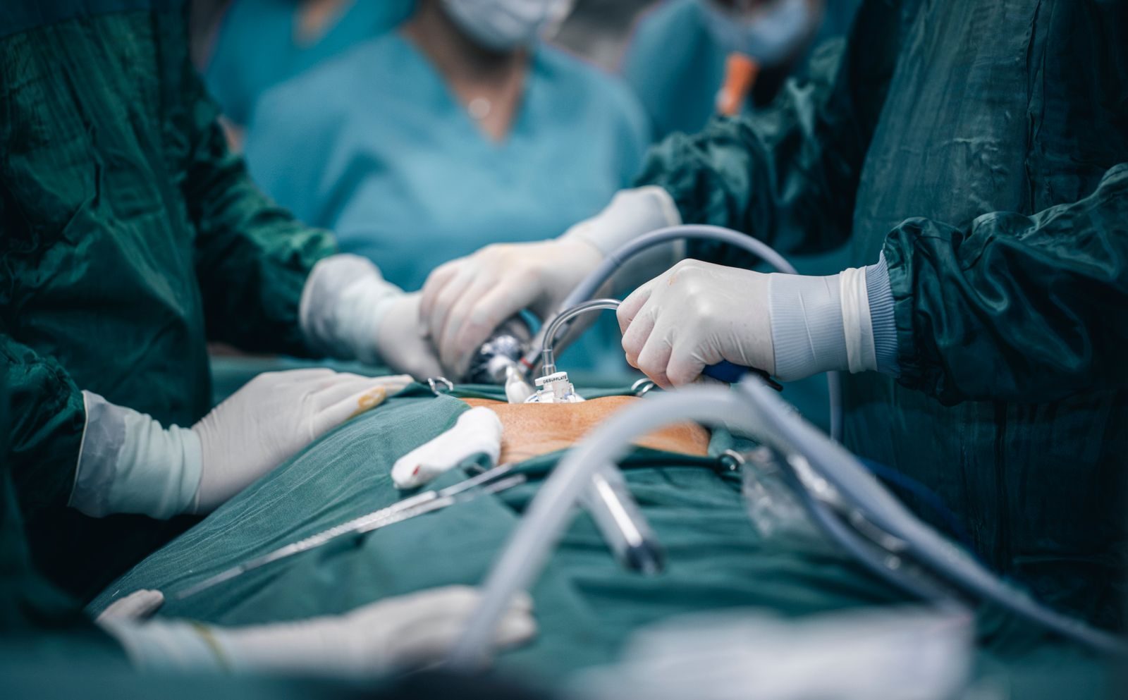
The clearance of a camera based exoscope for open surgery adds a new tool to the surgical toolkit. In June 2026, the FDA approved Karl Storz’s 4K Rubina Lens exoscope for use in open procedures. This digital camera system projects a magnified view of the surgical field onto a monitor, giving the surgeon and entire team the same high definition perspective. When surgeons switch to near infrared (NIR) mode after injecting indocyanine green (ICG) dye, the Rubina Lens highlights blood flow, lymphatic channels and tissue perfusion in real time, revealing critical details that could significantly change how surgical decisions are made.
Why It Matters
Intraoperative visualization is one of the few things surgeons can change instantly to improve outcomes. Traditional surgery depends on the surgeon’s direct view or a microscope, but an exoscope delivers that view to a high resolution screen. Now the whole team sees the same image, which improves coordination and teaching. When the view switches to fluorescence after ICG injection, it adds functional information. For example, it can show whether a suture line in bowel surgery has strong blood supply or not.
Operating rooms are busy, costly environments and a new device shifts workflow and priorities. Teams must learn to integrate the exoscope ,For example the timing of the dye injection, positioning the camera and toggling modes at key moments. Initially this may make cases run a little longer as staff adapt. Over time, however, the extra clarity could save time and uncertainty. For instance, if fluorescence confirms good perfusion, surgeons might avoid extra cutting or rechecking tissue; if it shows a problem, they can fix it immediately.
Beyond immediate decisions, this technology could enhance quality control and education. The exoscope records its high definition images, creating a video record of blood flow and lymphatic mapping. Surgeons can review these videos after the operation, use them in teaching conferences, or include them in the medical record. Over time, teams may develop guidelines about what counts as “adequate” fluorescence in various tissues. Professional groups are already studying best practices. For example, recent GI surgery guidelines recommend using ICG to confirm blood flow in intestinal anastomoses.
Who It Affects
Patients stand to benefit if this tool works as intended. Anyone undergoing surgery where blood flow or tissue viability is a concern could see improvements. This includes patients having bowel resections, organ transplants, cancer operations or reconstructive flap procedures. For example, confirming a well perfused gut anastomosis may reduce leak rates and complications. Identifying key lymph nodes during cancer surgery could improve tumor removal. In short, patients may experience fewer complications and faster recoveries.
The surgeons and the surgical team members will experience it firsthand. With the use of the exoscope, there is a shift in how the surgeons perform their work; from having to bend over the deep cavity, they will have to operate using the monitor, sometimes in a more convenient position (most surgeons experience less strain on their necks as a result). The assistants, the nurse, and even the anesthesiologist will see the same thing on the monitor, which increases the coordination among the people in the operating room.
Leaders and payers at hospitals need to consider cost vs. benefit analyses. Exoscope technology is an investment that is similar in nature to sophisticated imaging equipment in the operating room. It depends upon the benefits of this technology to offset costs, as even a couple of expensive complications avoided by using the technology would make the investment worth it. Otherwise, it would be difficult for hospital management to justify the cost of the equipment.
The regulators and professional organizations will give direction. If the FDA clears the technology, then it is deemed to be safe for use for its intended purposes, but not necessarily a recommendation on how to use it. The surgical organizations will evaluate the literature and come up with recommended guidelines. For instance, they can recommend that high-risk patients be routinely evaluated for blood flow to the anastomosis or flap perfusion through the use of ICG. In the future, this skill may be part of the training curriculum and board examinations.
What Changes
- Real-time fluorescence imaging in open surgery becomes routine. Surgeons can visualize blood flow and lymphatic drainage directly in the open field. For example, if the planned edge of an intestinal resection shows poor dye uptake, the surgeon can trim more tissue until only well-perfused areas remain. Decisions about cuts, anastomoses, and flap placement will increasingly incorporate this immediate feedback, potentially reducing guesswork.
- Team-based visualization becomes the norm. With the exoscope projecting a 4K image, everyone in the operating room sees the same detailed view. This enhances coordination and education. If a consultant is called into the case, they can instantly see the situation on screen. Trainees benefit greatly by observing each step on the monitor rather than peeking over shoulders. The team’s situational awareness improves when the operative field is literally in view for all.
- Workflow and training will shift. Staff will learn new steps: timing the ICG injection, placing the camera, and recognizing fluorescence patterns. The team may pause briefly after injecting ICG to watch the first wave of fluorescence. Surgeons will train to identify normal versus abnormal perfusion patterns – for example, knowing what a healthy tissue glow looks like. There will be a learning curve: initial cases may go slower, but with experience, toggling modes and interpreting images will become second nature.
- Procurement and interoperability are important. Hospitals already using digital OR platforms (such as Karl Storz IMAGE1 systems) may add the exoscope with minimal fuss. Others might need new monitors, mounts, or integration with existing video systems. Technical teams will ensure the exoscope’s output works with displays and recording devices. If multiple vendors are involved, adapters or additional equipment may be required. These logistical choices affect both the cost and the speed of implementation.
- Clear advantages emerge with the exoscope. Surgeons can work comfortably without straining their bodies, and the entire procedure can be recorded in 4K. This high-quality recording provides a precise surgical log for documentation and teaching. The exoscope also “democratizes” the surgeon’s view: instead of only the person at the eyepiece seeing the field, the whole team sees it on screen. In practice, this means adding fluorescence capability into the surgery without needing a separate bulky camera for fluorescence.
- Limitations and cautions must be acknowledged. Fluorescence is not a foolproof test. Bright fluorescence typically indicates good blood flow, but a dim or absent signal doesn’t always mean failure – sometimes tissue may still survive on collateral vessels. Ambient OR lights, blood, or technical issues can also obscure the image. Timing is key: looking too early or late after dye injection can mislead. In practice, surgeons must combine the camera’s information with traditional checks, like feeling pulses or seeing tissue directly. In other words, it should complement but not replace clinical judgment.
- Cost and access are challenges. As with many new medical devices, exoscopes will likely appear first in well-funded academic and urban hospitals. If the Rubina Lens proves to significantly improve outcomes, demand will grow and prices may stabilize. Until then, smaller or rural hospitals may not have it. This gap could widen inequity if patient care improves with the technology. Healthcare systems and policymakers may need to consider strategies – such as shared equipment or referral centers – if this technology truly helps patients.
- Looking ahead, surgeons and researchers will explore this technology’s impact. Early adopters may publish cases describing situations where fluorescence changed the plan (for example, prompting extra tissue removal or confirming anastomosis safety). Over time, formal studies and trials will likely measure outcomes like complication rates or healing times. The technology will also evolve: future software might quantify perfusion intensity, assigning a numeric blood flow score – shifting it from subjective judgment to a more data-driven assessment.
- Broader ecosystem factors will influence success. The exoscope is most useful when its images integrate smoothly into hospital systems. Ideally, surgeons could save fluorescence videos in the patient’s electronic record or a research database. That would allow teams across the hospital – or even between institutions – to review and learn from each case. However, if the system uses proprietary formats that don’t play well with others, sharing may be limited. Adopting standard video formats and linking operative images to patient records can help ensure these videos become shared learning tools.
- Adoption will ultimately depend on demonstrated benefit. Hospital leaders will want evidence: does using the exoscope actually reduce complications, shorten stays, or improve outcomes? Surgeons will want it to be reliable and intuitive. Industry, clinicians, and payers will need to collaborate on training programs and usage guidelines. If research shows a clear advantage – for instance, significantly fewer reoperations – adoption will accelerate. If benefits remain uncertain, it may stay limited to research centers or specialty hospitals.
- The FDA clearance is an important milestone, but it’s just a first step. It gives surgeons a powerful new way to see the surgical field, but how that changes patient care depends on people. If surgical teams embrace this extra information and integrate it thoughtfully, it could improve precision and safety in the OR. If not, the Rubina Lens might remain just another gadget. Only clinical experience and data will tell whether this technology truly becomes “what matters” in the operating room.
References
- U.S. Food and Drug Administration. K254242 – KARL STORZ ICG Imaging System with RUBINA® Lens. FDA 510(k) decision letter. 2026. View FDA decision letter [\[accessdata.fda.gov\]](https://www.accessdata.fda.gov/cdrh_docs/pdf25/K254242.pdf)
- Society of American Gastrointestinal and Endoscopic Surgeons. 2025 Guidelines for Fluorescence Image-guided Surgery Using Indocyanine Green in Gastrointestinal Procedures. SAGES. 2025. View guideline [\[sages.org\]](https://www.sages.org/publications/guidelines/figs-with-icg-guidelines/)
- Esposito C, Lepore B, Cerulo M, et al. Applications of indocyanine green (ICG) fluorescence technology in open surgery: preliminary experience in pediatric surgery. Frontiers in Surgery. 2023. View article [\[frontiersin.org\]](https://www.frontiersin.org/journals/surgery/articles/10.3389/fsurg.2023.1238487/full)
- Cho SS, Teng CW, De Ravin E, et al. Assessment and Comparison of Three Dimensional Exoscopes for Near-Infrared Fluorescence-Guided Surgery Using Second-Window Indocyanine Green. Journal of Korean Neurosurgical Society. 2022. View article [\[ncbi.nlm.nih.gov\]](https://www.ncbi.nlm.nih.gov/pmc/articles/PMC9271809/)
One story a day
The story of the day, in your inbox
One health journey each morning — no advice, no alarm, just company for the road.