Imaging May Extend Stroke Thrombolysis to 24 Hours
Selected patients with salvageable brain tissue may benefit from IV thrombolysis 4.5 to 24 hours after ischemic stroke onset, although symptomatic intracranial hemorrhage risk is higher.
 Written and medically reviewed byDr. Abu BakarContributing writer · PharmD, PhD (Pharmacology)
August 13, 2026 · 6 min read

A wider window, but only for selected patients
A 2026 systematic review found that intravenous thrombolysis given 4.5 to 24 hours after acute ischemic stroke improved 90-day functional outcomes among patients selected with advanced imaging. The benefit did not apply to patients chosen by time alone. Participants had imaging patterns intended to identify viable, hypoperfused brain tissue that might still be saved despite the delayed presentation.
The tradeoff was clinically important: symptomatic intracranial hemorrhage occurred more often with thrombolysis than with control treatment. That signal means an extended treatment window should not be interpreted as routine eligibility for everyone arriving within 24 hours. It describes a narrower population in whom the prospect of preserving threatened tissue may outweigh the additional bleeding risk.
The review synthesized randomized evidence rather than relying solely on observational comparisons, strengthening the inference that treatment contributed to the functional difference within the populations studied. However, the supplied PubMed record should be consulted for the exact number of included trials, pooled population, effect estimates, confidence intervals and statistical definitions. These details should not be reconstructed from secondary summaries when treatment protocols or policy decisions are being considered.
The outcome of greatest practical relevance was function at 90 days, generally assessed with the modified Rankin Scale. Late thrombolysis shifted outcomes in a favorable direction among imaging-selected patients, while the safety analysis showed more symptomatic intracranial bleeding. The evidence therefore supports a benefit–risk assessment, not a simple extension of the conventional clock-based window.
How imaging changes eligibility
The conventional 4.5-hour window uses elapsed time as a rough surrogate for tissue viability. Advanced imaging asks a different question: is there still threatened but salvageable brain surrounding a smaller region of established infarction?
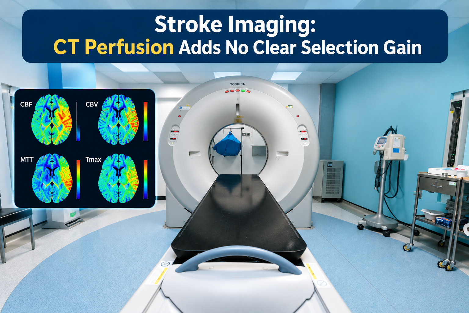
Computed tomography perfusion or magnetic resonance imaging can estimate the irreversibly injured core and the larger area with delayed or reduced blood flow. A meaningful mismatch between these regions suggests that some tissue remains at risk rather than already infarcted. MRI diffusion and fluid-attenuated inversion recovery mismatch may also help select patients whose onset is unknown, including wake-up stroke, although that approach is not identical to perfusion selection out to 24 hours.
Late-window trial populations were highly selected. In broad terms, they included adults with a clinically meaningful ischemic deficit, no intracranial hemorrhage, a limited established infarct and imaging evidence of salvageable tissue. Individual trials differed in vessel status, imaging software, mismatch thresholds, stroke severity, age restrictions, time windows and thrombolytic agent. Those criteria should not be treated as interchangeable.
Selection also requires the usual assessment for contraindications to thrombolysis, including active bleeding, relevant recent procedures, severe uncontrolled hypertension, anticoagulant exposure and laboratory abnormalities when applicable. Advanced imaging does not neutralize those risks. Nor does a favorable mismatch establish that treatment will benefit a particular patient.
Where thrombectomy fits
For an eligible patient with a treatable large-vessel occlusion, mechanical thrombectomy remains central to late-window reperfusion care. Evidence for imaging-selected thrombolysis does not justify delaying vascular imaging, endovascular consultation or transfer to a thrombectomy-capable center.
The new evidence is most relevant when thrombectomy is not readily available, cannot be performed within a clinically useful period, is contraindicated, or is not indicated because no accessible large-vessel occlusion is present. In those circumstances, a hospital with rapid perfusion imaging and established stroke expertise may have an additional reperfusion option for a carefully selected patient who would otherwise receive supportive treatment alone.
“Unavailable” should be defined operationally rather than assumed from geography. A transfer pathway may still provide timely thrombectomy, and intravenous treatment should not become a reason to abandon that pathway. Local protocols need to specify who reviews the imaging, which mismatch criteria are accepted, how hemorrhage risk is assessed and whether treatment can begin without delaying transfer.
The choice of thrombolytic also matters. Trials across the extended-window literature have evaluated alteplase, tenecteplase or both in different clinical settings. Their findings cannot automatically be pooled into a single drug protocol, and the evidence does not support inventing a dose or substituting one agent for another outside the relevant trial criteria, labeling and institutional guidance.
The hemorrhage signal deserves equal weight
Symptomatic intracranial hemorrhage is uncommon but potentially devastating. Its increase in the systematic review is not a technical footnote; it is the principal counterweight to the better distribution of 90-day functional outcomes.
Risk may be influenced by infarct size, blood pressure, age, glucose, stroke severity, cerebral small-vessel disease, anticoagulant exposure and treatment delay. Imaging can exclude a large established core or visible hemorrhage, but it cannot eliminate bleeding risk. Definitions of symptomatic hemorrhage also vary across trials, which can complicate comparisons and pooled estimates.
At a policy level, extending thrombolysis requires more than acquiring perfusion software. Hospitals need reliable image acquisition, validated processing, rapid specialist interpretation, blood-pressure management, post-thrombolysis monitoring and immediate access to repeat brain imaging and hemorrhage treatment. A protocol that increases treatment access without those safeguards may not reproduce trial outcomes.
Shared decision-making remains difficult because stroke treatment is time-sensitive and patients may lack capacity. When feasible, clinicians can explain that imaging suggests living tissue remains at risk, that thrombolysis may improve the chance of less disability, and that it also raises the chance of serious intracranial bleeding. The evidence informs that discussion but does not predict an individual outcome.
Important uncertainties
The review inherits differences among its included trials. Imaging definitions, treatment windows, thrombolytic agents, vessel occlusion patterns, control groups and hemorrhage definitions may vary. Statistical pooling can estimate an average effect, but it does not prove that every imaging-selected subgroup has the same benefit or risk.
Generalizability is another concern. Trial centers commonly have experienced stroke teams, rapid imaging workflows and strict eligibility review. Results may differ in hospitals where perfusion studies are delayed, image quality is inconsistent or specialist interpretation is unavailable. Patients with large established infarcts, severe baseline disability or other major bleeding risks may also have been underrepresented or excluded.
The most useful next evidence would clarify which mismatch thresholds best predict net benefit, whether outcomes differ between alteplase and tenecteplase, how vessel status modifies response, and how late thrombolysis performs when transfer for thrombectomy is possible but substantially delayed. Cost-effectiveness and implementation studies are also needed for smaller and rural hospitals.
Questions clinicians ask
Does every patient presenting within 24 hours need perfusion imaging?
No. Imaging should answer a clinically relevant eligibility question and should not delay standard treatment for patients already within an established window. In later or unknown-onset presentations, perfusion imaging or an appropriate MRI mismatch approach may identify salvageable tissue, but local capability and the criteria used in the supporting trial matter.
Can thrombolysis replace thrombectomy for a large-vessel occlusion?
Not when timely thrombectomy is feasible and the patient is eligible. Late-window thrombolysis may be considered when thrombectomy is unavailable, substantially delayed, contraindicated or not anatomically indicated, but treatment should not postpone vascular imaging, endovascular consultation or transfer to an appropriate stroke center.
How should the bleeding signal affect treatment decisions?
It should be considered alongside the potential improvement in 90-day function. A favorable perfusion mismatch does not erase hemorrhage risk, so clinicians still need to assess infarct burden, blood pressure, anticoagulants, recent procedures and other contraindications while using the eligibility and safety definitions from the applicable evidence.
Are alteplase and tenecteplase interchangeable in this window?
The extended-window literature includes different agents, populations and imaging criteria. Evidence from one regimen should not be transferred automatically to another, and this review does not support an improvised dose. Drug selection should follow the specific evidence, applicable US guidance and a hospital’s reviewed stroke protocol.
References
- IV thrombolysis beyond 4.5 hours improved 90-day outcomes in selected stroke patients — PubMed, 2026
- Thrombolysis Guided by Perfusion Imaging up to 9 Hours after Onset of Stroke — The New England Journal of Medicine, 2019
- MRI-Guided Thrombolysis for Stroke with Unknown Time of Onset — The New England Journal of Medicine, 2018
- Guidelines for the Early Management of Patients With Acute Ischemic Stroke: 2019 Update — Stroke, 2019
One story a day
The story of the day, in your inbox
One health journey each morning — no advice, no alarm, just company for the road.