Stroke Imaging: CT Perfusion Adds No Clear Selection Gain
Pooled evidence found no significant difference in functional independence or symptomatic intracerebral hemorrhage when CT perfusion and noncontrast CT were used to select patients for acute ischemic stroke treatment.
 Written and medically reviewed byDr. Abu BakarContributing writer · PharmD, PhD (Pharmacology)
September 20, 2026 · 6 min read
TheBrief
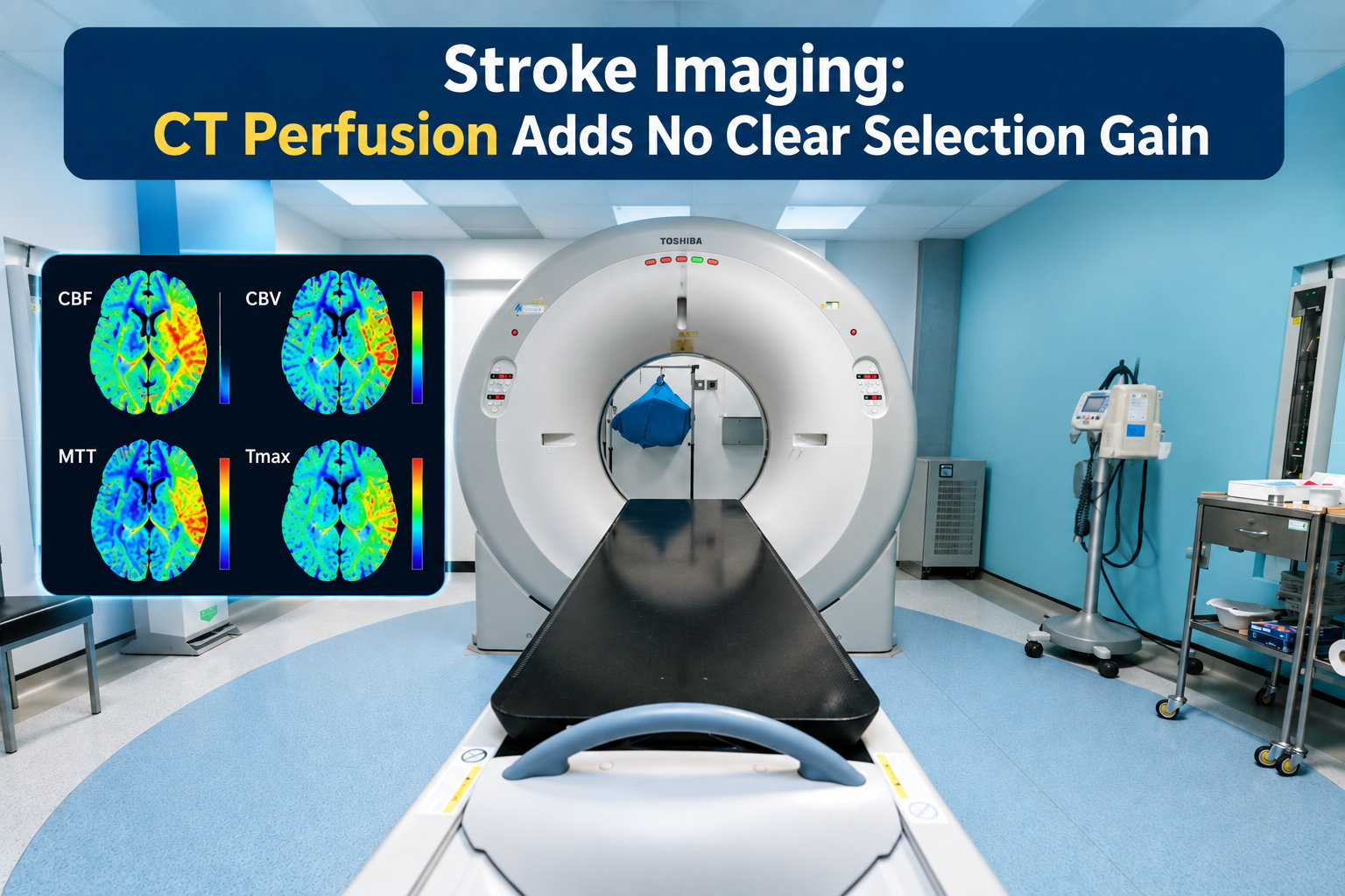
CT perfusion has not shown a clear improvement in functional independence or symptomatic intracranial hemorrhage outcomes compared with noncontrast CT-based selection in the pooled evidence used to inform the 2026 stroke guideline process. The finding does not mean CT perfusion has no role. Instead, it suggests that its added acquisition, processing and transfer demands should have a clear clinical purpose, particularly when every minute can affect time-sensitive stroke treatment.
No measurable outcome advantage in pooled data
The systematic review and meta-analysis informing the 2026 American Heart Association/American Stroke Association guideline process found no statistically significant difference between CT perfusion-based and noncontrast CT-based selection in the two outcomes most relevant to the comparison: functional independence and symptomatic intracerebral hemorrhage. Functional outcome was assessed using the modified Rankin Scale, generally at 90 days in the underlying acute stroke literature.
The finding is narrower than saying CT perfusion has no clinical value. It indicates that, across the comparative data pooled by the investigators, adding perfusion imaging as a selection tool did not translate into a detectable increase in the proportion of patients achieving functional independence. Nor did it produce a detectable difference in symptomatic bleeding risk.
These are group-level findings, not proof that the modalities are interchangeable in every presentation. CT perfusion estimates ischemic core and hypoperfused tissue, while noncontrast CT primarily identifies hemorrhage and established ischemic change. The technologies answer different imaging questions even when downstream outcomes appear similar.
What the comparison actually tested
The review synthesized studies comparing treatment selection strategies built around CT perfusion with strategies using noncontrast CT. The population was adults evaluated for treatment of acute ischemic stroke, particularly reperfusion therapy. The analysis was based on study-level comparative evidence rather than a randomized assignment of patients to one imaging workflow or the other.
That design matters. Imaging modality is often determined by hospital capability, clinician preference, treatment window, local transfer pathways and the severity or clarity of the presenting scan. Patients sent for perfusion imaging may therefore differ systematically from those selected with simpler imaging. Meta-analysis can increase precision, but it cannot eliminate confounding already present in the component studies.
“Noncontrast CT selection” also should not be read as “noncontrast CT alone in all cases.” In contemporary thrombectomy pathways, noncontrast CT is commonly paired with CT angiography to identify a large-vessel occlusion and evaluate vascular anatomy. The practical comparison is often a streamlined CT-plus-angiography pathway versus a pathway that adds perfusion acquisition and software processing.
The pooled absence of a statistically significant difference is not evidence of exact equivalence. A confidence interval crossing the null means that the analysis did not establish superiority or harm; it does not exclude smaller benefits or disadvantages. Interpretation should account for the precision and heterogeneity reported in the source analysis rather than relying only on the direction of the point estimate.
Speed, access and transfer logistics
The principal implication is operational. If CT perfusion does not provide a demonstrated outcome advantage for the average patient represented in the pooled evidence, systems should avoid allowing it to become an automatic barrier to reperfusion treatment. This is especially relevant at primary stroke centers, rural hospitals and facilities that lack around-the-clock perfusion software or specialist interpretation.
Every added imaging step has a potential opportunity cost. Acquisition, image transfer, automated processing, quality review and repeat scanning after technical failure can delay thrombolysis, transfer or arterial access. The size of that delay varies substantially by institution. A mature comprehensive stroke center may complete perfusion imaging without meaningful interruption, while a transferring hospital may lose critical time waiting for processing or remote review.
Noncontrast CT remains essential for excluding intracranial hemorrhage and estimating the extent of early ischemic injury. CT angiography can then establish whether a treatable large-vessel occlusion is present. Where these studies provide sufficient information to make a treatment or transfer decision, the meta-analysis offers no outcome-based reason to require perfusion imaging routinely.
CT perfusion may still add selection value when conventional imaging leaves genuine uncertainty. Examples include an unclear time of onset, discordance between clinical severity and visible infarction, concern about a large established core, or a need to characterize potentially salvageable tissue in an extended treatment window. Its role is strongest when the result is likely to change a decision rather than merely document physiology already apparent from the examination, noncontrast CT and angiography.
Randomized thrombectomy trials provide important context but do not settle the modality comparison. SELECT2, for example, established a benefit from endovascular thrombectomy in selected patients with large ischemic cores, using eligibility criteria that could incorporate noncontrast CT ASPECTS or estimates from advanced imaging. Such trials support treating appropriately selected patients; they do not show that one imaging modality produces better outcomes than another selection pathway.
For policy, the evidence favors capability-based protocols. Hospitals with rapid, reliable perfusion imaging can retain it for defined indications. Hospitals without it should not necessarily delay transfer to obtain it elsewhere when hemorrhage has been excluded, a large-vessel occlusion has been identified and the receiving center can complete any additional assessment. Protocol performance should be evaluated using treatment and transfer times, rates of technically inadequate scans, treatment eligibility and patient outcomes—not imaging sophistication alone.
Important limitations and unanswered questions
The principal limitation is nonrandomized modality selection in much of the evidence base. Confounding by indication, differences in stroke severity, treatment window, occlusion site, collateral status and local expertise can influence both the choice of imaging and the outcome. Definitions of symptomatic intracerebral hemorrhage, perfusion thresholds and favorable functional outcome may also differ across studies.
CT perfusion is not a single standardized intervention. Scanner hardware, contrast timing, motion correction, vendor algorithms and thresholds for ischemic core or penumbra can produce materially different estimates. A pooled comparison may obscure strong performance in optimized programs and weaker performance where acquisition or processing is less reliable.
Generalizability also remains uncertain for patients at the margins of trial and cohort eligibility, including those with very large infarcts, posterior-circulation occlusions, severe preexisting disability or unusually long transfer times. More useful future studies would randomize imaging strategies within treatment windows and report workflow intervals, transfers avoided or delayed, treatment rates, functional outcomes, hemorrhage and resource use.
The present evidence therefore supports restraint rather than abandonment. CT perfusion can answer clinically useful questions, but pooled outcomes do not justify treating it as a universal gatekeeper when simpler imaging can support a timely decision.
Questions clinicians ask
Can thrombectomy selection proceed without CT perfusion?
Yes, in appropriate pathways. Noncontrast CT can exclude hemorrhage and assess early ischemic change, while CT angiography can confirm a large-vessel occlusion. The pooled evidence did not show better functional independence with routine CT perfusion-based selection, although individual eligibility still depends on the treatment window, clinical findings and local protocol.
Does this mean CT perfusion should be skipped in late-window stroke?
No. Advanced imaging may clarify tissue viability when onset is unknown or conventional imaging is inconclusive. The evidence supports using it selectively when the result could alter treatment, while avoiding delays when noncontrast CT, angiography and the clinical picture already provide enough information for a treatment or transfer decision.
Is noncontrast CT alone sufficient before transfer?
It may be sufficient to begin an urgent referral, but vascular imaging is usually important when large-vessel occlusion is suspected. A streamlined noncontrast CT and CT angiography protocol can provide the key information needed by a thrombectomy center without requiring the referring hospital to perform perfusion imaging.
How should stroke systems apply these findings?
Systems should measure whether perfusion imaging changes decisions and how it affects door-to-treatment and transfer times. Routine use is harder to justify where processing is slow or unreliable; selective use is more defensible when rapid acquisition is available and the result resolves a specific uncertainty.
References
1. Response to Request for Modification of the 2026 AHA/ASA ... — PubMed Central, 2026 2. Guidelines for the Early Management of Patients With Acute Ischemic Stroke: 2019 Update to the 2018 Guidelines — American Heart Association/American Stroke Association, 2019 3. Trial of Endovascular Thrombectomy for Large Ischemic Strokes — The New England Journal of Medicine, 2023
One story a day
The story of the day, in your inbox
One health journey each morning — no advice, no alarm, just company for the road.